


Heart failure can be considered a veritable epidemic due to its prevalence. In France, it affects around 1.5 million people, a figure that is set to rise in line with the aging population.
Due to the diversity of its causes, this condition affects all areas and subspecialties of cardiology and cardiac surgery. A multidisciplinary approach is essential to greatly improve the quality of life and life expectancy of patients. Drug treatments, pacemakers and defibrillators, percutaneous approaches, and surgery were addressed and discussed during this International Day.
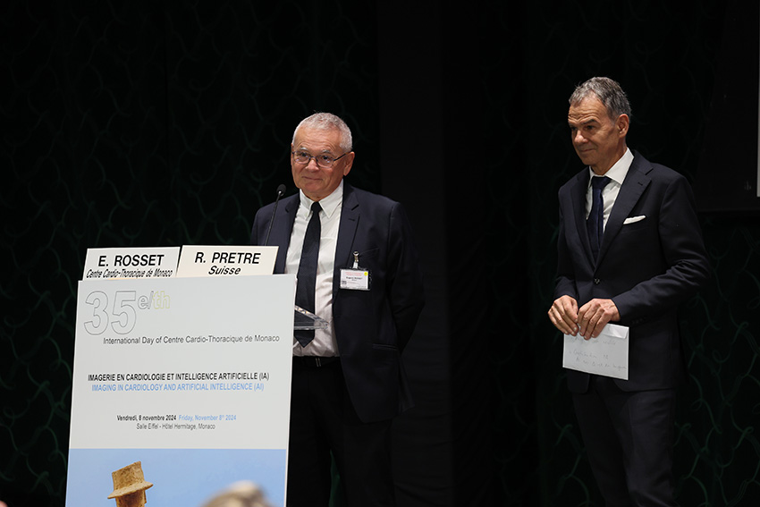
At the end of this enriching Day, Professor René Prêtre presented “The Fabulous History of Heart Transplantation,” an epic worthy of the best Hollywood screenplays, and Luc Ferry presented “The Symbolism of the Heart, Between Philosophy, Science, and Mythology.”
International Day organized by the Scientific Council of the Monaco Cardio-Thoracic Center on the theme of “Aortic aneurysms: state of the art”.
Internationally renowned experts provided a detailed overview of aortic aneurysms. The epidemiology of aortic aneurysms (frequency, incidence, risk factors, location, evolutionary risks, including rupture) is crucial to a better understanding of these conditions, and to improving their prevention, screening and treatment. A full update was also given on current recommendations for pre- and post-operative monitoring, and medical and surgical treatment of aneurysms.
Detailed presentations on the endovascular aortic repair in a variety of aneurysms, including the most complex, the diagnosis and treatment of endoleaks, the place of laparoscopic or robot-assisted aortic surgery, and an update on AI and digital twin technologies in the planning of aortic stents, were the highlights of this International Day.
A meeting of excellence in the image of the CCM, which has built its reputation for over 35 years on its medical-surgical expertise in the management of cardiac and vascular pathologies.
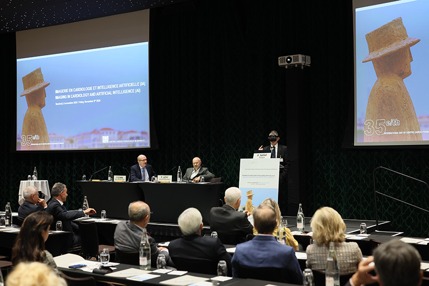
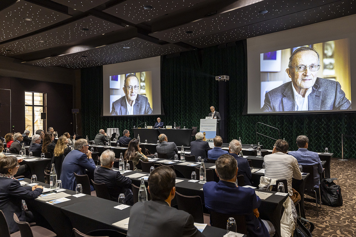
On Friday 8th November 2024, the 35th International Day of the Monaco Cardio-Thoracic Centre was held in the Salle Eiffel at the Hôtel Hermitage.
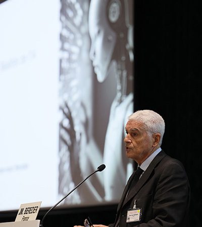
The use and contribution of artificial intelligence in cardiology is progressing rapidly. European experts took stock of the use of A.I. today in the treatment of cardiovascular pathologies, particularly in imaging, which is essential in interventional cardiology and cardiac and vascular surgery.
The benefits of using A.I., its contribution to diagnosis and treatment, its most promising applications, its potential impact on the training of cardiologists, but also its current limitations, were the highlights of this International Day.
It was a meeting of excellence that reflected the image of the CCM, whose reputation has been built on its medical and surgical expertise for over 35 years.
Watch the Monaco Info story – Le 19h – Edition of Friday 8th November 2024
From heart attack to recovery: Bruno’s journey through advanced cardiac care
The Cardio-Thoracic Centre is proud to have taken part in this project, which has enabled us to demonstrate how the hospital and the Principality are organized to deliver a complementary and effective range of care.
Pr. Sylvie DI FILIPPO Monaco Cardiothoracic Center.
Pediatric and adult congenital cardiology & fetal cardiology.
University professor, hospital practitioner.
Congenital heart disease is the most common congenital organ anomaly. Their incidence in the general population is 14 foetuses per 1000 and 8 births per 1000.
Congenital heart disease includes multiple malformations of varying anatomical and functional complexity and with heterogeneous prognoses.
These malformations are classified according to their severity and complexity into:
Minor, moderate and major
Repairable and non-repairable
Biventricular and univentricular
Cyanogenic and non-cyanogenic
Minor heart disease
No need for intervention
Moderate heart disease
Intervention indicated
Repairable heart disease
Severe heart disease
Heart disease requiring treatment and intervention
Repairable with sequelae or non-repairable
Bicuspid aortic valve
Small atrial septal defect
Small interventricular septal defect
Small ductus arteriosus
Minor mitral valve anomaly
Minor pulmonary narrowing
Pulmonary vein anomalies
Coronary anomalies
Aortic stenosis
Atrioventricular canal
Large AIC
Large IVC
Large ductus arteriosus
Aortic coarctation
Tight pulmonary stenosis
Marfan’s disease and aortic aneurysm
Ebstein’s tricuspid anomaly
Tetralogy of Fallot
Transposition of large vessels after repair
Heart disease with fixed pulmonary hypertension (Eisenmenger)
Non-operated or palliated cyanotic heart disease
Interruption of the aortic arch
Right ventricle with double outlet
Single ventricles
Pulmonary atresia
Common arterial trunk
Univentricular Fontan circulation
Transposition of the great vessels with right ventricle under the aorta
Complex heart disease with multiple lesions
Anatomo-physiopathological classification of congenital heart disease
Left-right shunts
Right-to-left shunts and right obstructions
Left obstructions
Coronary anomalies
Rhythmological abnormalities
Complex heart disease
Shunt: natural or created abnormal communication between heart chambers or vessels.
Cyanosis: blue discolouration of the skin and mucous membranes due to a drop in oxygen in the blood (or hypoxia).
Antenatal screening
Congenital heart disease is well tolerated during intrauterine life and does not prevent normal development of the foetus.
Adaptation to extra-uterine life, which involves a fall in pulmonary pressures and closure of the ductus arteriosus and foramen ovale, is the cause of decompensation of cardiac malformations, the viability of which depends on the permeability of these shunts.
Antenatal diagnosis of heart disease by echocardiography and Doppler enables decompensation to be anticipated and prevented, by guiding immediate post-natal therapeutic management. The birth is programmed and planned by the gynaeco-obstetric and neonatology team.
In the majority of cases, pre-term induction is not necessary. Only cases of fetal heart failure may justify it (the consequence of an abnormal heart rhythm, either too slow = fetal bradycardia, or too fast = uncontrolled fetal tachycardia).
Antenatal screening enables heart disease to be diagnosed, with results ranging from 40% to 90% depending on the type of heart disease.
It enables a diagnosis to be made, the course of the disease to be monitored antenatally, postnatal care to be planned and the prognosis to be established.
Birth planned in this way helps prevent decompensation of the heart disease after birth
The prostaglandin E1 administered is a treatment that keeps the ductus arteriosus open at birth, thereby maintaining circulation and/or oxygenation in newborns with heart disease at risk of acute decompensation.
Heart diseases at risk of neonatal decompensation entail a lethal risk, including::
– Right-sided obstructions that limit pulmonary flow
– Left-sided obstructions that limit aortic flow
– Mixing anomalies that depend on the patency of the foramen ovale
Diagnosis and clinical symptoms
Newborn
Cyanosis, heart failure
Shunt-dependent heart disease
Symptoms
Feeding difficulties
Poor growth or weight stagnation
Frequent bronchopulmonary infections
Dyspnoea on exertion
Feeling unwell, syncope
Chest pain
Asymptomatic
Breathing
ECG abnormality discovered by chance
Abnormalities on clinical examination: femoral pulses, high blood pressure
Auscultation: detection of a murmurInfant weight growth curve: weight stagnation (red dots)
Cardiac and Doppler ultrasound: cardiac ultrasound is a simple, non-invasive examination that can be used to diagnose cardiac malformations at any age.
Neonatology
Screening for congenital heart disease
Antenatal
heart disease detected antenatally must be confirmed postnatally by a cardiac ultrasound scan of the newborn.
Postnatally
not all heart disease is detected before birth, and it is still very important to examine all newborn babies in the maternity unit to look for any heart abnormality that may have gone unrecognised, and to recognise it before the baby is discharged home.
The clinical examination should look for :
palpation of the femoral pulses
The presence of a murmur on ausculation
The newborn’s feeding behaviour
Oxygen saturation or satO2: systematic screening for heart disease in newborns is carried out in maternity units by measuring oxygen saturation in the upper and lower limbs. The normal level is > 97%, with a difference of less than 3% between the upper and lower limbs. If this measurement is abnormal, an echocardiogram is performed to check for any cardiac abnormality before the baby is discharged from the maternity hospital.
Saturation sensor
Imaging congenital heart disease
IRM
Magnetic resonance imaging makes a major contribution to the assessment of congenital heart disease, particularly in adulthood, when the performance of echocardiography is less good. MRI provides additional information that is essential for assessment, follow-up, diagnosis, management and therapeutic decisions. It allows :
Anatomical assessment of cavities and vessels
Functional assessment: of myocardial function
Analysis of flow and output
Search for myocardial fibrosis
Testing for ischaemia using the adenosine test
Its use is limited in children because of the need for sedation
The main indications are systemic right ventricles (double mismatch, transposition of the great vessels with atrial switch), right ventricles with volume overload (tetralogy of Fallot), single ventricles, pulmonary, aortic, tricuspid and mitral valve disease, aortic pathologies, etc.
SCANNER
The angio-scanner is a complementary imaging technique which allows the vessels to be viewed in particular:
pulmonary arteries
pulmonary veins
coronary arteries
aorta
veno-venous and arteriovenous fistulas
Its use is limited by irradiation and injection of potentially nephrotoxic and allergenic contrast medium.
Congenital heart disease in adulthood
Over 90% of children with congenital heart disease (CHD) survive into adulthood.
The number of adults with CHD is now greater than the number of children.
Most of these patients cannot be considered cured, and their management is a lifelong process: from repair in childhood, to the transition to adulthood, to the desire for pregnancy, and the management of late complications specific to each CC.
ESC 2020 recommendations: management of congenital heart disease in adulthood.
Transition to adulthood
The transition to adulthood is an important phase that needs to be anticipated and supported, so that patients can receive all the information they need about their heart disease, their medical history, their treatment, their follow-up and all the problems associated with their heart disease.
To this end, therapeutic education programmes have been developed and set up in the referral centres.
At the CCM: the transition is made naturally by an organisation based on a medical-surgical team caring for the same patients from foetus to adulthood.
Complications of congenital heart disease in adulthood:
Myocardial dysfunction: left ventricle, single ventricle, systemic right ventricle
Coronary artery disease
Heart failure
Arrhythmias
Infectious endocarditis
Arterial hypertension
Transmission and genetics of congenital heart disease
Embryology of the normal heart: the heart is formed during the first 8 weeks of foetal life, in several successive complex stages starting from the primitive cardiac tube, with phenomena of torsion and rotation and partitioning:
Primitive heart tube
Loop: position of the atria/ventricles cavities
Formation of the atrioventricular valves
Partitioning of the atria
Partitioning of the ventricles
Partitioning of the vessels
Congenital heart disease is inherited through a multifactorial mechanism involving both genetic and environmental factors.
In the majority of cases (72%), no genetic aetiology is identifiable.
Genetic and environmental factors: account for 20% to 30% of cases
Environmental factors: 2% of cases
Maternal illnesses: diabetes, rubella, systemic lupus erythematosus.
Maternal use of treatments such as lithium, isotretinoin and anticonvulsants.
Maternal age: this is a risk factor for trisomy 21, which can lead to heart malformations.
Chromosomal abnormalities (aneuploidies): 10%.
trisomy 21 or Down syndrome, trisomy 18, trisomy 13, monosomy X or Turner syndrome
monogenic: congenital syndromes affecting multiple organs
Di George syndrome (microdeletion 22q11.2)
Williams-Beuren syndrome (microdeletion 7p11.23)
Single gene defects: fibrillin-1 mutations (Marfan syndrome), TXB5 (Holt-Oram syndrome)
PTPN11 (Noonan syndrome).
Autosomal dominant de novo point mutation: 8%.
Autosomal recessive inherited point mutation: 2%.
The risk of recurrence of congenital heart disease in a family depends on the cause: negligible in de novo mutations, 2 to 5% in non-syndromic multifactorial congenital heart disease, and 50% when an autosomal dominant mutation is involved.
It is important to identify genetic factors in order to assess the risk of recurrence and to guide preconception genetic counselling.
Table: examples of genetic mutations with associated heart disease.
How can we help you?
Gérer le consentement
Pour offrir les meilleures expériences, nous utilisons des technologies telles que les cookies pour stocker et/ou accéder aux informations des appareils. Le fait de consentir à ces technologies nous permettra de traiter des données telles que le comportement de navigation ou les ID uniques sur ce site. Le fait de ne pas consentir ou de retirer son consentement peut avoir un effet négatif sur certaines caractéristiques et fonctions.
Fonctionnel
Always active
Technical access or storage is strictly necessary for the legitimate purpose of enabling the use of a specific service explicitly requested by the subscriber or user, or for the sole purpose of carrying out the transmission of a communication over an electronic communications network.
Preferences
Technical access or storage is necessary for the legitimate purpose of storing preferences that are not requested by the subscriber or user.
Statistics
Technical storage or access that is used exclusively for statistical purposes.Technical storage or access that is used exclusively for anonymous statistical purposes. Without a subpoena, voluntary compliance on the part of your Internet Service Provider, or additional records from a third party, information stored or retrieved for this purpose alone cannot usually be used to identify you.
Marketing
Technical access or storage is necessary to create user profiles for the purpose of sending advertisements, or to track the user on a website or across multiple websites with similar marketing purposes.
Contact +377 92 16 80 00 Emergencies relating to cardiovascular and thoracic pathology are accepted without restriction, 24 hours a day, 7 days a week, Sundays and public holidays included.