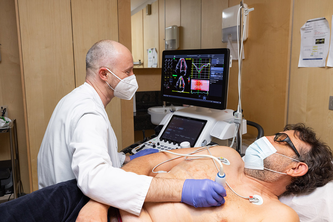
For over 30 years, the Monaco Cardiothoracic Centre has built its international reputation for medical and surgical expertise using latest generation technical equipment. This innovative specificity is based on a partnership with one of the world’s major players in medical technology, Siemens Healthineers.
The CCM: Siemens Healthineers’ first reference centre
For more than 20 years, the CCM and Siemens Healthineers have established a strategic partnership in cardiovascular medicine, creating innovative solutions to treat patients suffering from cardiovascular disease.
In 2010, the CCM became the first Siemens Healthineers Reference Centre for cardiovascular disease. The original purpose of this collaboration was to share skills (training of practitioners by the CCM) and to guide development towards new solutions (clinical advice, validation of new diagnostic concepts.)
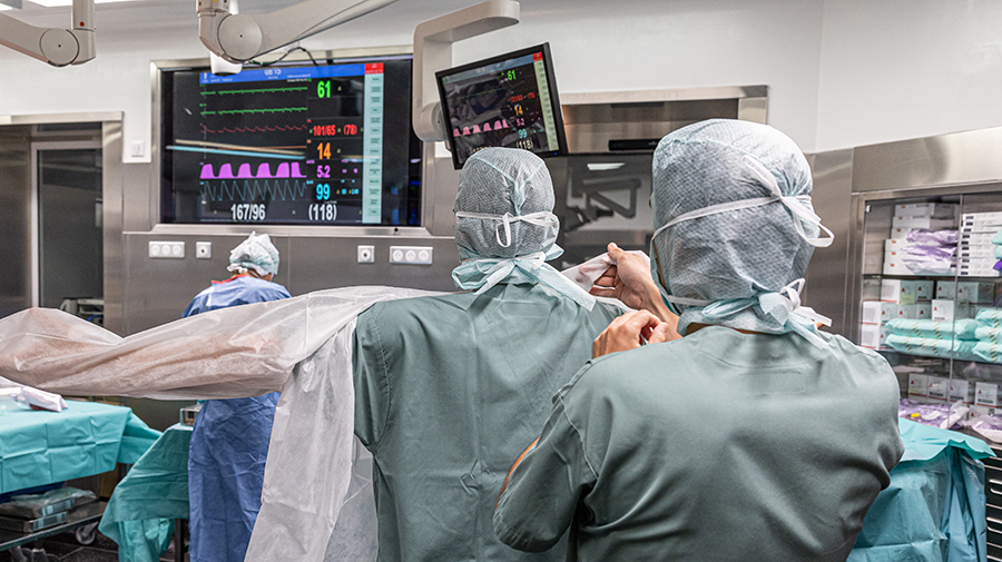
The CCM’s technology and its latest generation medical imaging tools enable patients to be treated in a comprehensive manner in a structure where treatment can be carried out simultaneously and collegially in the same location.
All the specialists (clinical & medical imaging doctors, anaesthetists / intensive care doctors, surgeons) work together in the same one-stop shop (reception, hospitalization, paramedical staff, examination and imaging rooms, intensive care rooms and operating theaters). This single location allows patients to be treated in a collegial environment, offering the best care without needing to transfer patients or duplicating examinations, also avoiding the existence and financing of five different departments.
Hybrid rooms
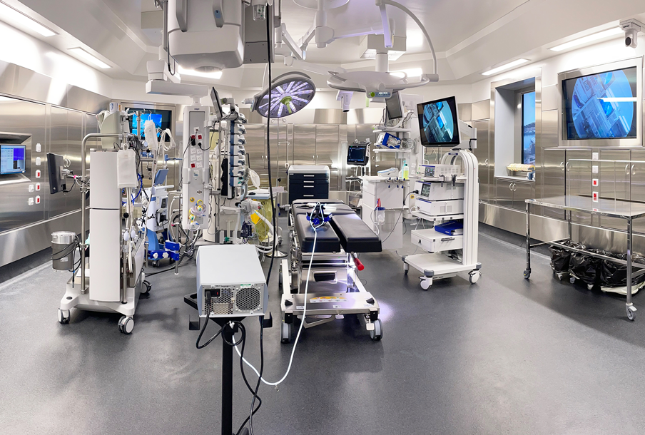
The CCM has 2 hybrid rooms equipped with the latest Siemens Healthineers’ interventional systems. Equipped with a fully robotic C-arm, the hybrid rooms are equipped with technological tools that allow the integration of scanner and ultrasound imaging by merging them with radiological images.
They have been completely re-equipped recently, notably with the new generation of ARTIS Pheno and ARTIS Icono systems.
These rooms thus optimize minimally invasive therapeutic management of patients in cardiac surgery, interventional cardiology and vascular surgery.
Operating theaters
Designed to facilitate and secure the most delicate interventions, the two operating theaters are spacious (70_m2 each), ergonomic, bright and meet strict access and organization standards.
Access to the theaters via an airlock-room with rigorous hygiene and complete clothing and shoe change for all the medical staff on each occasion they enter.
Particular attention has been paid to the separation of the septic/cleansing circuits: peripheral circulation eliminates undesirable crossing.
The non-use of external blood in extracorporeal circulations is privileged in the Centre since its opening. This, together with all aseptic, hygienic and maintenance measures that are implemented on the 4th floor and hospitalisation floors, has enabled the control of the risk of infection since the first years of operation.
- Dräger anaesthesia workstations.
- Getinge operation tables
- LivaNova extracorporeal circulation pumps.
- Dräger monitors.
- Dräger lighting.
High-tech imaging equipment is permanently available for cardiothoracic and vascular diseases, thus eliminating any delay or time lapse between the need for an examination and its completion.
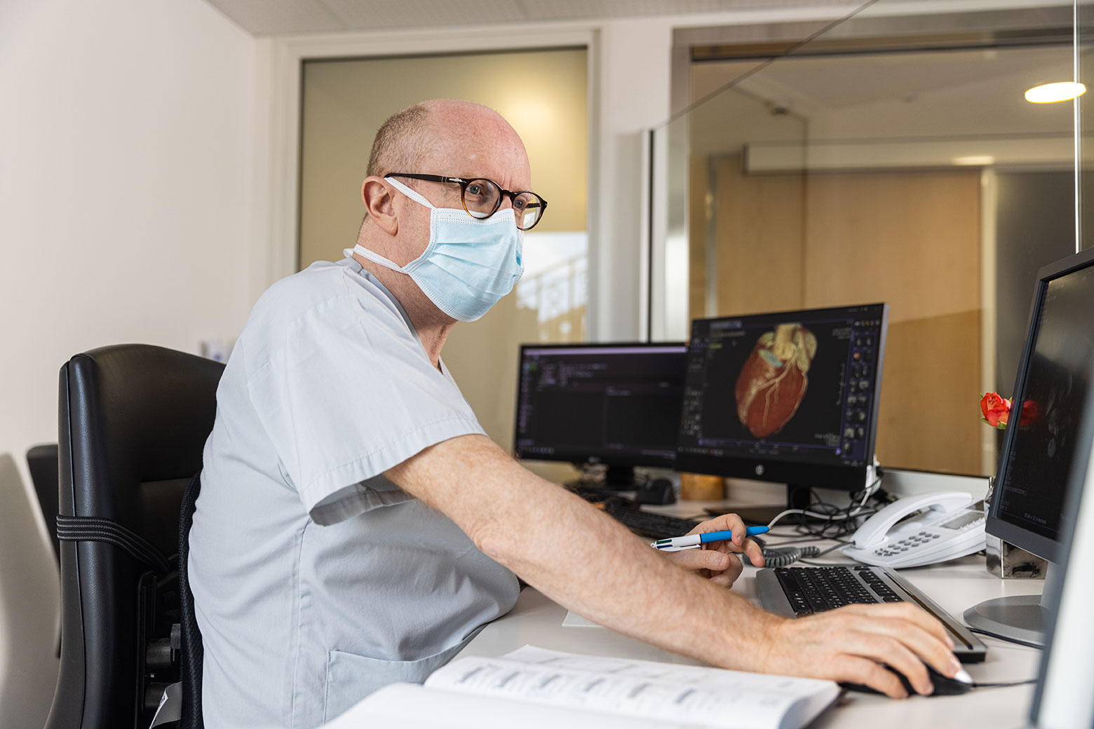
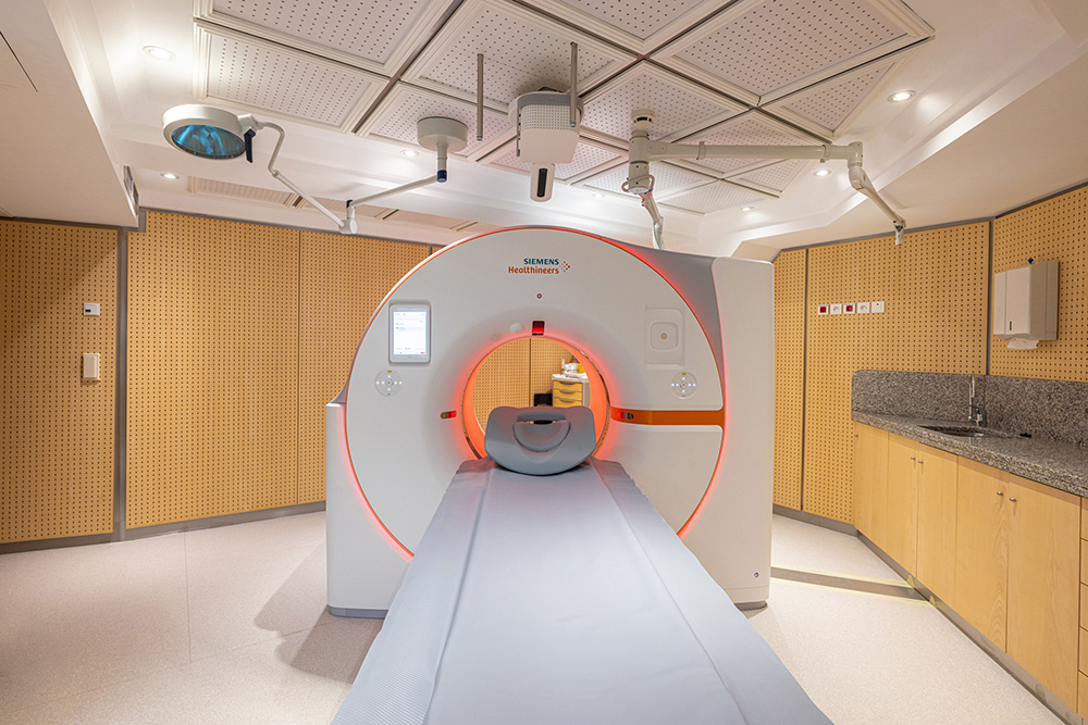
As part of its partnership with Siemens Healthineers, the Cardio-Thoracic Centre was the first private centre in the world to be equipped with a photon-counting scanner dedicated exclusively to cardiovascular pathologies, the Naeotom Alpha.
This latest-generation scanner represents a technological revolution in medical imaging and a major step forward in patient care. The high precision of its images means that it can be used to improve diagnostic capabilities, enabling patients to be treated more effectively or to benefit from early treatment.
Cardiologists performing CT examinations :
- Dr Filippo CIVAIA: +377 92 16 82 19
- Dr Philippe ROSSI: +377 92 16 82 19
For more information, download the explanatory sheet.
MRI or Magnetic Resonance Imaging allows images of the human body to be taken using a magnetic field (magnet) in which radiofrequency waves pass.
These waves put into “resonance”, that is to say that they vibrate the hydrogen atoms that make up the tissues of the body, and thus produce images.
MRI does not use X-rays.
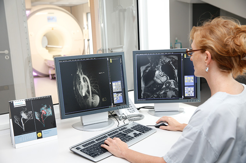
Cardiac MRI allows the morphology and function of the heart muscle and valves to be analysed and detect, for example, a contraction defect in the heart muscle, the narrowing of a valve, etc.
Vascular MRI allows the analysis of large and small vessels: thoracic or abdominal aorta, supra aortic trunk, renal arteries, the arteries of the lower limbs.
Cardiologists performing MRI examinations :
- Dr Filippo CIVAIA: +377 92 16 82 19
- Dr Laura IACUZIO: +377 92 16 82 19
For more information, download the explanatory sheet.
The peculiarity of cardiac MRI with Adenosine
This examination allows heart exploration (heart valves and cavities).
Cardiologists carrying out echocardiograms :
- Dr Franck LEVY: +377 92 16 82 92
Exercise tolerance (stress) test
Dobutamine stress echocardiogram
Transesophageal Echocardiogram
